Chemical imaging: Antibiotic interaction on individual bacterial cells with unprecedented resolution
- Light
- Life
Published: | By: Dr. D. Täuber
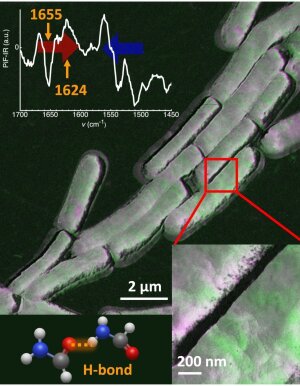
Cover Art The interaction of the antibiotic vancomycin on the surface of individual Bacillus subtilis cells, visualized using mid-infrared photoinduced force microscopy (PiF-IR).
Image: Maryam Ali, Dr. Daniela TäuberMid-infrared photoinduced force microscopy (PiF-IR) is a new imaging technique that enables the chemical characterization of surfaces with an unprecedented resolution of less than 5 nm. PiF-IR bridges the gap between high-resolution structure elucidation using electron microscopy, fluorescence microscopy and conventional infrared spectroscopy. It is complementary to tip-enhanced Raman spectroscopy (TERS).
Chemical imaging of bacterial surface with less than 5 nm resolution
An interdisciplinary team of researchers led by Dr. Daniela Täuber in Jena has successfully mapped the local chemistry and structure of the antibiotic interaction on the surface of individual bacterial cells with a resolution of a few nanometers using the model system Bacillus subtilis and vancomycin. Their work demonstrates the potential of this novel infrared spectroscopy imaging method for applications in biomedical research and has been published in Analytical Chemistry: “Nano-chemical cell-surface evaluation in photothermal spectroscopic imaging of antimicrobial interaction in model system Bacillus subtilis & vancomycin” (doi:10.1021/acs.analchem.5c03502External link). The high spatial and spectral resolution of PiF-IR in particular enables the visualization of chemical changes on the surface of pathogens that result from a local interaction with antibiotic substances.
Findings from collaborative project pintXsum
The data analysis approach developed by the team is largely based on findings on hybrid field coupling in nanostructured surfaces from the project "pintXsum" (Photo-INduced Thermal eXpansion of rough SUrface Morphologies). This collaborative project by PD Dr. Christin David (Institute of Condensed Matter Theory and Optics) and Dr. Daniela Täuber (Institute of Physical Chemistry) was funded by the FSU for development within the profile lines LIGHT and LIFE. They combined PiF-IR measurements on a polymer nanoparticle with modeling its photothermal expansion. From this, they could attribute the frequently observed anisotropic signal distributions on soft nanostructures in tip-enhanced photothermal imaging methods to hybrid field coupling; see S. Anindo, D. Täuber, C. David: Photothermal Expansion of Nanostructures in Photo-induced Force Microscopy, published in the Journal of Physical Chemistry (doi:10.1021/acs.jpcc.4c08305External link). So far, such field coupling has been known for metallic nanostructures mainly.
Successful collaboration in LIGHT and LIFE
More than 150 years after the pioneering work of Abbe, Zeiss and Schott in the field of light microscopy, the fruitful collaboration between theoretical physics, experimental microscopy and biomedical applications in microscopy still provides new and fundamental approaches to biomedical knowledge!
-
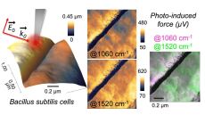
Mid-infrared photoinduced force microscopy (PiF-IR) enables nanochemical imaging of a bacterial surface.
Image: © 2025 The Authors. Publ. by AmChemSoc (publ. is lic. under CC-BY 4.0) -
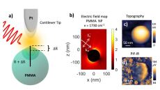
Hybrid coupling of the incident electric field between a platinum-coated AFM tip and a polymer nanosphere. Model and experiment using mid-infrared photoinduced force microscopy (PiF-IR).
Image: © 2025 The Authors. Publ. by AmChemSoc (publ. is lic. under CC-BY 4.0)