Next generation microscopy methods - Ghost imaging
Forschungsthema geleitet von Zhe Sun
This research project is supported by Deutsche Forschungsgemeinschaft (DFG, German Research Foundation) under Germany´s Excellence Strategy – EXC 2051 [Project-ID 390713860], “Balance of the Microverse”. This project aims for a novel method of high-resolution, low-dose, and label-free microscopy, enabling different and/or new views on biological specimens including cells and their extracellular fluid as well as for cytoplasm and organelles. We will explore the applicability of high-quality microscopic ghost imaging based on laboratory light sources in the extreme ultraviolet (XUV) spectral region. Microscopy with short wavelength radiation ensures few nanometer resolution and element-specific absorption without any additional labeling. microscopic ghost imaging on the other hand warrants high contrast and high-resolution microscopy with a dose well below the radiation damage level.
Ghost imaging, which is an unconventional imaging technique developed in the frame of quantum imaging and relies on quantum or spatial intensity-fluctuation correlations to image objects, will allow us to overcome some of these limitations. Both entangled photon interference and classical intensity-fluctuation correlations could be used for ghost imaging. Ghost images are obtained by correlating the total intensity of the transmitted or reflected light of an illuminated object with the spatially resolved intensity of a position-correlated reference beam that has never interacted with the object. Therefore, radiation damage of the sample can be alleviated by varying the intensity ratios between object- and reference-beam, which is the most severe limitation for enhancing the resolution in classical biological nanoscale imaging.
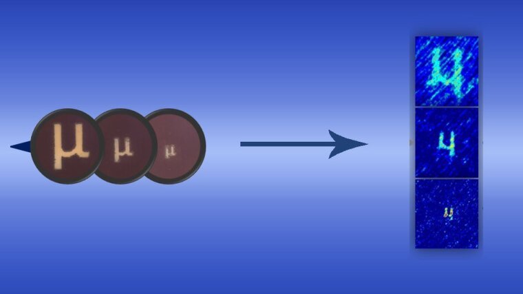
The principle of pseudo-thermal ghost imaging is shown in Fig.1. A He-Ne laser and a rotating diffuser constituted a pseudo-thermal light source. The rotating diffuser is in the focal plane of the lens with a focus lens. The generated speckle field is divided by a beam splitter into two spatially correlated beams, namely the transmitted (reference path) and the reflected (object path) beam. The object beam hits the object “μ” and the so-called bucket signal is collected with a photodiode. The reference beam never interacts with the object and is detected by the photodiode at the same distance with a CCD camera. Pseudothermal ghost imaging relies on correlating a measured 2-D intensity speckle pattern to a transmission or reflection scalar value of an object illuminated by the same speckle pattern. The randomness of the rotating ground-glass diffuser in the pseudothermal ghost imaging will impose the field fluctuations that arise. By varying the speckle pattern over time and simultaneously measuring the transmitted light through the object, a statistical reconstruction of the image is possible, see Fig.2. The retrieved image quality directly depends on the number of iterations and the correlation between fluctuating patterns and bucket signals.
Pseudo-thermal ghost imaging has great potential for nanoscale microscopic imaging, especially for high-resolution XUV ghost imaging using a table-top XUV light source, where ghost imaging could be an alternative technique for radiation-sensitive samples like cells. XUV microscopic ghost imaging technique is expected to become an important tool for imaging with low flux.
We are welcome for complete internships, bachelor, and master thesis within the project.
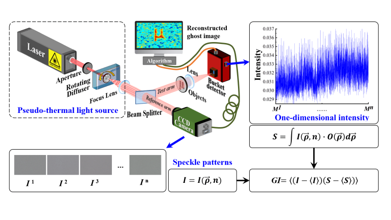
Fig. 1. Schematic of pseudothermal ghost imaging.
Abbildung: Zhe SunThe collimated laser beam through a rotating ground-glass diffuser. The beam splitter copies the speckle patterns into the reference and object beams through the same distance free-space paths. The reference beam illuminates a high-spatial-resolution detector (CCD camera) whereas the test beam illuminates a single-pixel detector (bucket detector) through object transparency. I=I(ς,n) is the intensity distribution of the signal speckle field. S is the total light intensity containing the object’s information.O(ς) is the objective function and ς=(x,y,z) is the space coordinate. n is the total number of iterations. The ghost image is retrieved by GI=[(I-[I])(S-[S])] .
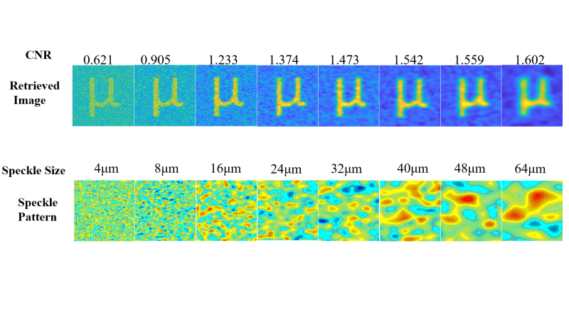
Fig. 2. The contrast of noise ratio of the retrieved ghost image vs speckle size.
Abbildung: Zhe Sun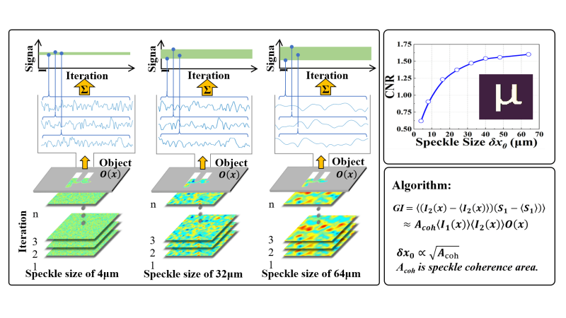
Fig. 2. The contrast of noise ratio of the retrieved ghost image vs speckle size.
Abbildung: Zhe SunPublications: